Vertebra Lumbar Spine Anatomical Model - Degenerative Changes Disc Protrusions Disassemblable 3B Scientific
3B Scientific
Request a Quote
H2: Vertebra Anatomical Model
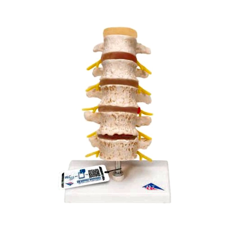
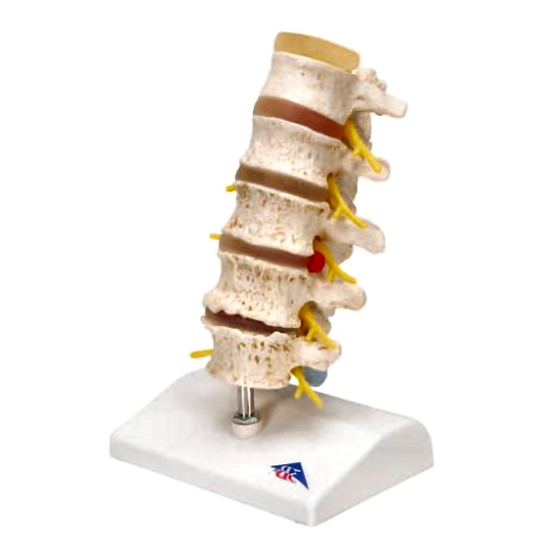
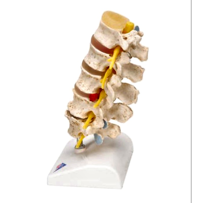
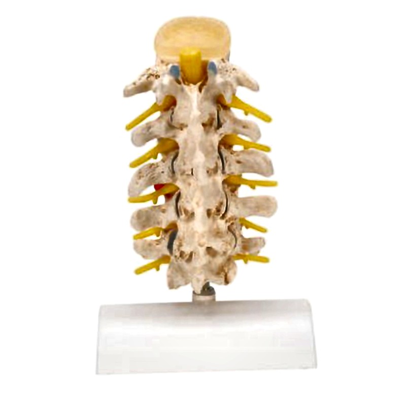
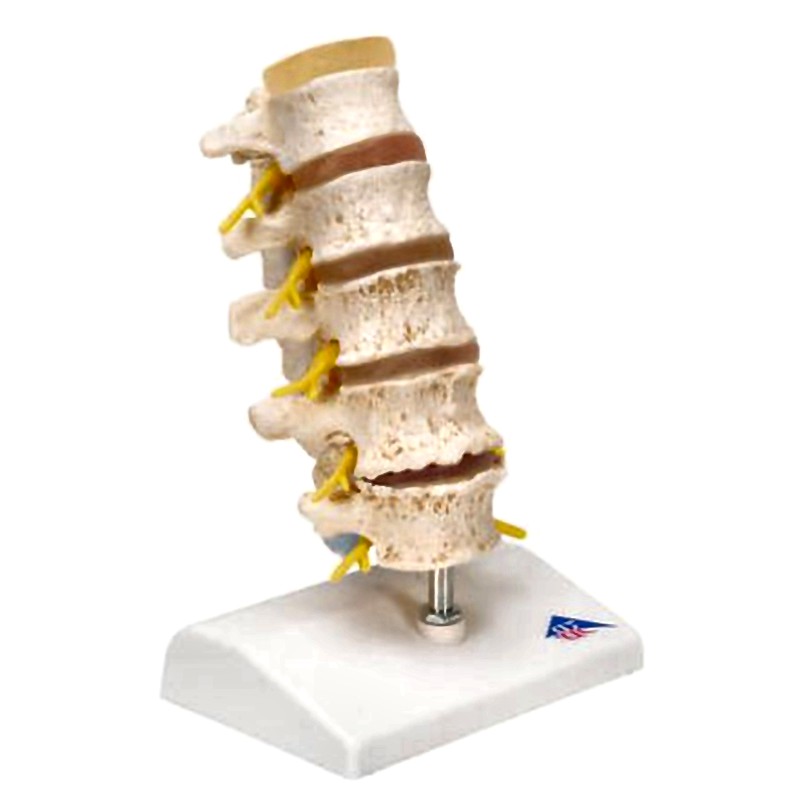
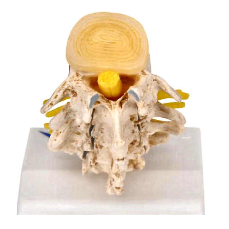
The model provides a detailed representation of degenerative changes affecting the vertebral bodies and intervertebral discs in the lumbar spine, illustrating conditions across a spectrum of severity. Based on an original cast of a human lumbar spine, fine bony structures are accurately reproduced to support advanced anatomical education.
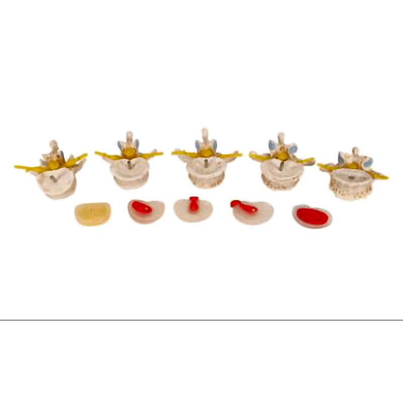
The sequence from top to bottom displays:
- Intervertebral disc in healthy condition
- Lumbar vertebra (L1) without degenerative changes
- Intervertebral disc with protrusion
- Lumbar vertebra (L2) with beginning degenerative changes
- Intervertebral disc with medial prolapse
- Lumbar vertebra (L3) with advanced degenerative changes
- Intervertebral disc with intraforaminal prolapse on the right
- Lumbar vertebra (L4) with strong degenerative changes and bony constriction of the left intervertebral foramen
- Extremely narrowed intervertebral disc
- Lumbar vertebra (L5) with strong degenerative changes and bony constriction of the left intervertebral foramen, demonstrating pressure on the spinal nerve root L5 on the left
The model is designed for disassembly into individual vertebrae and intervertebral discs to facilitate hands-on learning.
Supplied on a stable base for secure display and classroom use.
Every original 3B Scientific anatomy model now includes these additional free features:
- Free access to the 3B Smart Anatomy course, hosted within the award-winning Complete Anatomy app by 3D4Medical
- The 3B Smart Anatomy course contains 23 digital anatomy lectures, 117 interactive virtual anatomy models, and 39 anatomy quizzes for knowledge assessment
- Bonus: Free warranty upgrade from 3 to 5 years upon product registration
- Tip: A free 3-day trial to all premium features of the Complete Anatomy app is provided with enrollment in the 3B Smart Anatomy course
This model supports educational applications in anatomy, orthopedics, neurology, physical therapy, and spinal surgery training.